Dark Leaf Lesions: Fungal or Bacterial Disease?

 Dark leaf lesions usually signal disease, and you can often narrow the cause by key visual differences between pathogens. It explains how bacterial spots look, how fungal lesions develop and spread, plus simple testing clues for accurate ID. It also compares treatments by disease type and flags common misdiagnosis mistakes.
Dark, spreading leaf spots can make you wonder if the problem is fungal or bacterial. They may look alike at first, but they need different treatments, so guessing can waste valuable time and let damage spread. This guide helps you compare symptoms, check conditions, and pick a practical next step so you can act quickly and protect your plants.
What dark lesions usually indicate
When leaf spots turn brown, purple-black, or nearly so, it usually means the affected tissue is dying and the plant is trying to wall off the problem. That dark color can come from oxidized plant compounds, collapsed cells, and sometimes the pathogen’s own pigments. In practice, these marks often point to an active infection or a fast-moving stressor rather than a minor cosmetic issue.
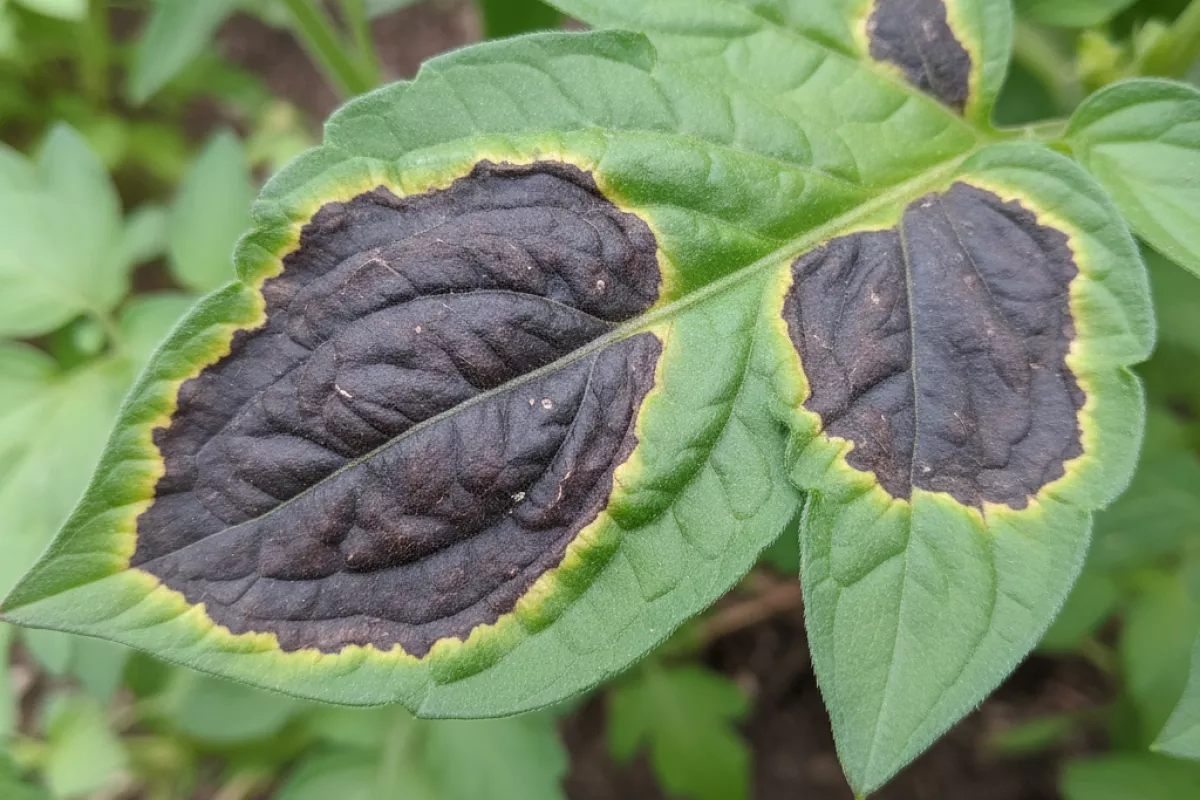
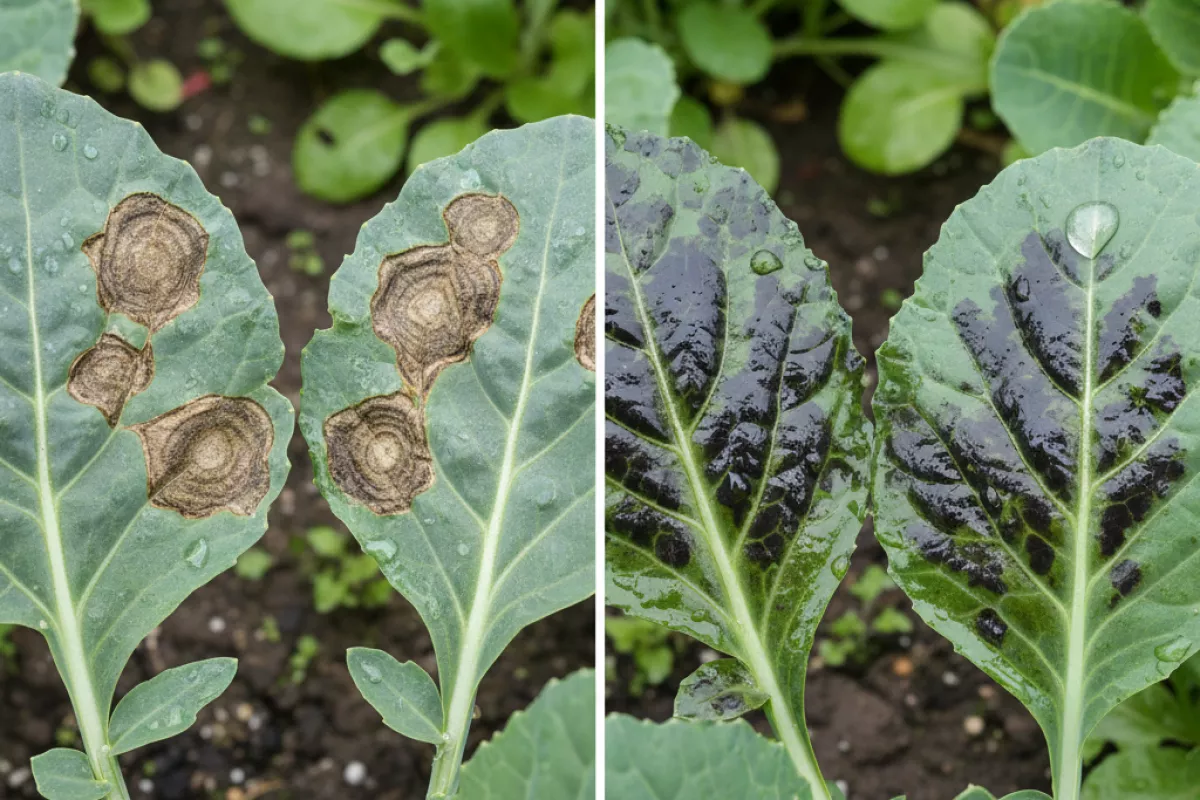
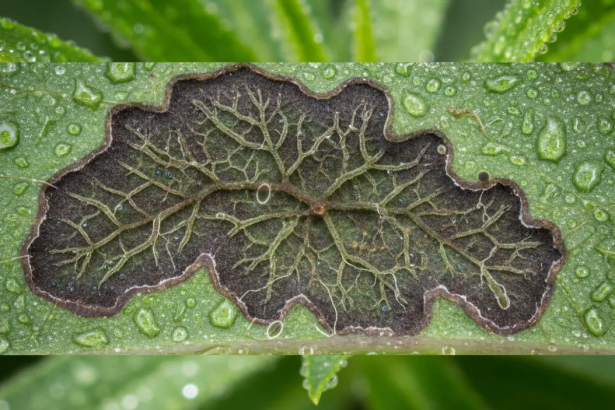
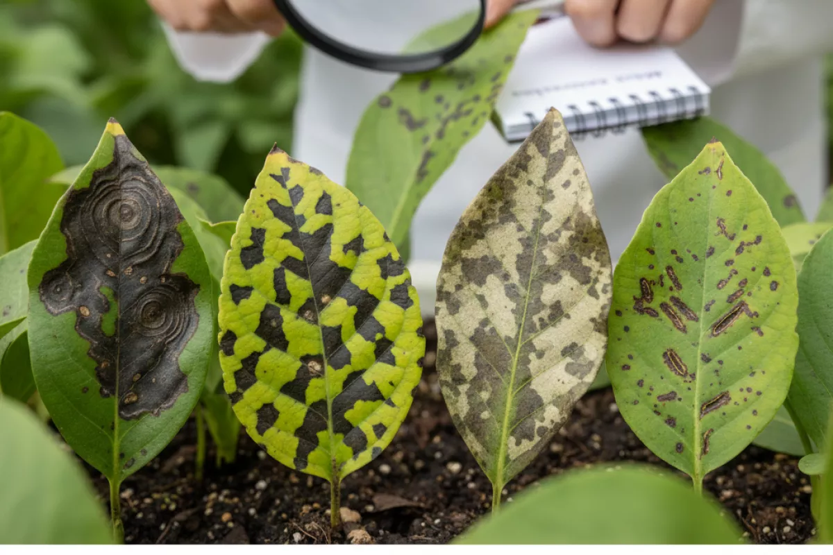
Darkened patches are most often associated with fungi or bacteria, but the pattern around the spot matters as much as the color. Some lesions stay sharply bordered and dry; others look water-soaked at first and then blacken. A yellow halo, a greasy sheen, or spots that “bleed” along veins can hint at bacteria, while concentric rings, fuzzy growth, or a more powdery/dry look tends to lean fungal. Still, there’s overlap, and the same plant can have more than one issue at once.
- Necrosis (dead tissue): Black or deep-brown centers typically mean cells have been killed, either by toxins from a pathogen or by the plant’s own defense response.
- Defense-related staining: Some plants darken tissue around an infection as they deposit phenolics and lignin-like compounds to slow spread.
- Moisture-driven spread: If spots start as water-soaked, then turn dark and expand after rain or overhead watering, bacteria becomes more plausible.
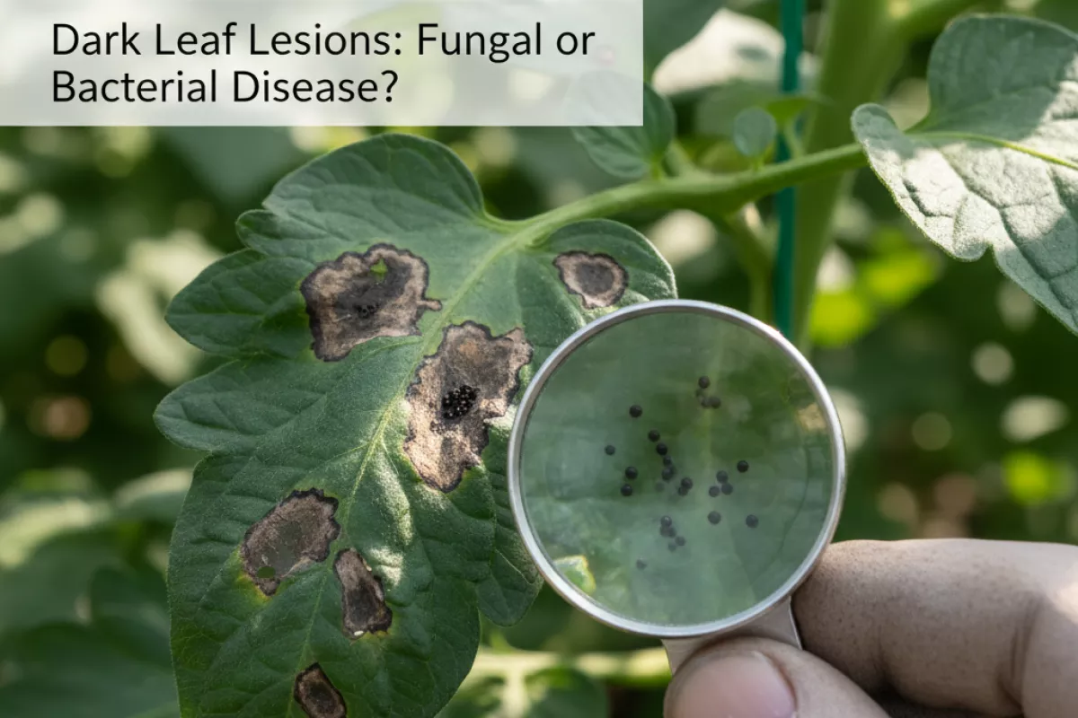
- Spore-producing infections: If you see tiny dark specks (like pepper) inside the lesion, that can be fungal fruiting bodies rather than “dirt.”
- Vein-limited patterns: Angular lesions that stop at veins often happen when microbes move through leaf spaces constrained by veins; this is common in bacterial leaf spot but not exclusive.
- Secondary colonization: Older damage from sunscald, chemical burn, or insect feeding can darken later as fungi move in, making the original cause easy to miss.
| Clue on the leaf | What it often suggests | Why it matters for next steps |
|---|---|---|
| Water-soaked spot that later turns dark; may look “greasy” | Bacterial involvement is more likely | Reduce leaf wetness (avoid overhead watering), sanitize tools, remove heavily affected leaves |
| Concentric rings (“target” pattern) or a dry, papery center | Fungal leaf spot is more likely | Improve airflow, avoid splashing soil, consider pruning for ventilation |
| Yellow halo around a dark center | Common with bacterial spots; also seen with some fungi | Look for additional signs (ooze, angular shape) before deciding on treatment |
| Angular lesions bounded by veins | Often bacterial, sometimes downy mildew-like infections | Monitor spread after wet weather; isolate plants if it’s moving quickly |
| Tiny black dots within the lesion | Fungal fruiting bodies are possible | Indicates the pathogen is reproducing; remove debris and infected leaves promptly |
| Dark scorch along edges or between veins without clear spots | Stress injury (salt, drought, fertilizer burn) or toxin exposure | Check watering and feeding practices before assuming disease is the primary cause |
A useful rule of thumb: if the lesion looks wet before it looks dark, suspect bacteria; if it looks dry and patterned (rings, specks, dusty growth), suspect fungi. Either way, dark leaf lesions usually mean the plant has already lost that tissue, so the goal shifts from “healing the spot” to slowing spread and correcting the conditions that let it take hold.
Key visual differences between pathogens
Start by looking at the shape of the spot, what happens at the leaf edge, and how the lesion behaves in wet vs. dry conditions. Fungal problems often build distinct patterns or textures on the surface, while bacterial issues more often look water-soaked and “greasy,” especially after rain, overhead watering, or heavy dew.
| What you see on the leaf | More typical of fungal leaf spots | More typical of bacterial leaf spots |
|---|---|---|
| Early lesion look | Dry-looking specks that expand; color shifts from tan/brown to dark, sometimes with a defined margin | Water-soaked, translucent patches that darken; often looks “oily” or wet at first |
| Spot shape and edge | Often round to oval with smoother borders; may show a crisp outline | Often angular because veins limit spread; edges can look jagged or vein-traced |
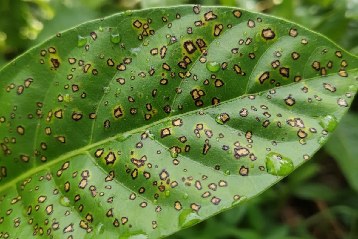
| Concentric rings (“target” pattern) | Common in several fungal diseases (rings or zonation as the spot expands) | Uncommon; lesions usually lack clear ringed pattern |
| Yellow halo around dark center | Can happen, but not always prominent | Very common; chlorotic halo can be wide and bright, especially on young leaves |
| Surface texture | May develop a dusty, velvety, or fuzzy coating (spores) in humid conditions | Usually no fuzz; surface stays smooth but may look shiny when wet |
| Lesion center | May dry out and crack; sometimes forms tiny black dots (fruiting bodies) | May turn papery and tear out, leaving “shot holes” on some plants |
| Pattern on the plant | Often starts on older, lower leaves and moves upward with splashing water and wind | Often appears after storms or overhead irrigation; can spread quickly through a blocky section |
| Response to humidity | Spots enlarge steadily; sporulation may appear after prolonged leaf wetness | Looks dramatically worse after a wet night; water-soaked margins are more obvious in the morning |
| Stem/fruit involvement | Some fungi cause sunken, dry cankers or scabby lesions | Some bacteria cause soft, wet rot or dark, greasy streaks; ooze may appear under very wet conditions |
- Do a “morning check.” If lesions look notably wetter or more translucent at dawn and then dry down by afternoon, that leans bacterial.
- Look for “targets” and tiny black pinpoints. Concentric rings or pepper-like dots in the center are classic fungal clues.
- Use a simple magnifier. With a 10× hand lens (about 0.4× in everyday terms), fuzzy growth or spore-like dust supports a fungal cause; a smooth, water-soaked look supports bacterial.
- Don’t rely on one sign. Dark leaf lesions can overlap visually, and mixed infections happen, so use several traits together before deciding on treatment.
How bacterial spots appear on leaves
Bacterial leaf problems usually show up as small, wet-looking marks that spread quickly after rain, overhead watering, or heavy dew. Early on, the tissue can look slightly translucent, as if it’s been water-soaked, before it turns brown or black. Because bacteria move in splashing water and enter through tiny wounds or natural openings, the pattern on the plant often looks more “scattered” than a slow, even creep from the edge.
As the spots mature, the plant’s response can create sharp borders, pale halos, or a yellow wash around the lesion. On some hosts, the dead center dries and becomes papery; on others, it stays greasy-looking. When infection pressure is high, individual lesions merge into larger irregular patches, and leaves may yellow and drop earlier than expected.
- Water-soaked start: fresh lesions often look dark, glossy, or translucent before they dry down.
- Angular shapes are common: spots may be limited by veins, creating blocky or “cornered” patches rather than perfect circles.
- Distinct margins: many bacterial lesions have a defined edge; a thin yellow halo can appear as surrounding cells react.
- Rapid change after wet weather: symptoms can seem to “explode” within 24–72 hours after warm, wet conditions (about 1–3 days).
- Shot-hole effect on some plants: dead tissue may crack and fall out, leaving small holes once the center dries.
- Streaking on stems or along veins: instead of round spots, you may see elongated, dark lines where bacteria track through tissue.
- Ooze under humidity: in very damp conditions, a sticky film or tiny beads can form on lesions; when dry, it may look like a thin crust.
A quick at-home check is to look at where the damage is concentrated. If the newest spots are clustered where water hits first (leaf tips, lower leaves splashed by soil, or areas under sprinklers), that supports a bacterial cause. Also note whether lesions stay sharply bounded by veins; that “angular” look is a frequent clue when you’re deciding between bacterial leaf spot and a fungal leaf lesion.
How fungal lesions develop and spread
Most dark spots caused by fungi begin when microscopic spores land on a leaf and find enough moisture to germinate. A thin film of water from rain, overhead irrigation, heavy dew, or high humidity gives the spore time to sprout and penetrate the cuticle, often through natural openings (stomata) or tiny wounds from wind, hail, or insects.
Once inside, the pathogen grows as threadlike hyphae between or within leaf cells and starts pulling nutrients from the tissue. That disruption is what you see as a lesion: cells collapse, pigments change, and the plant walls off the area. Many fungal leaf spots develop a distinct edge (sometimes with a yellow halo) and may show concentric rings or a target-like pattern as the infection advances in pulses.
- Spore arrival: Spores are carried by wind, splashing water, tools, hands, or infected transplants and debris.
- Germination window: Extended leaf wetness is the trigger; several hours of wet leaves can be enough, and repeated wet nights accelerate problems.
- Penetration and colonization: The fungus enters and spreads locally, killing small patches of tissue that merge into larger blotches.
- Reproduction on the leaf: New spores form on or in the dead tissue, often visible as tiny black dots, dusty growth, or velvety patches depending on the species.
- Secondary spread: Each wetting event can splash or wash fresh spores to nearby leaves, while breezes can move them farther within the canopy.
Spread is usually fastest where air circulation is poor and leaves stay wet: dense plantings, shaded beds, or plants watered from above late in the day. Warm conditions often speed fungal growth, but the exact temperature range depends on the pathogen; what matters most in real gardens is the combination of warmth plus repeated wet foliage.
| Stage in the leaf | What you may notice | What drives it |
|---|---|---|
| Early infection | Small, water-soaked flecks that darken; faint yellowing around the spot | Spore germination during long leaf-wet periods |
| Lesion expansion | Spots enlarge and may show rings or a defined margin; tissue may crack or become papery | Hyphal growth inside the leaf; plant tissue dies and dries |
| Spore production | Specks, dusty coating, or tiny black fruiting bodies in the dead center | Fungus reproduces on damaged tissue, especially after humidity spikes |
| Secondary spread | New spots appear higher up or on neighboring plants after rain or watering | Splash dispersal, wind movement, contact via hands/tools, contaminated debris |
Because many fungi can survive between seasons on fallen leaves or infected stems, outbreaks often restart from last year’s debris. That’s why the same patch of garden can see repeat dark leaf lesions even when the plant looks healthy early on: the first infections are small and easy to miss until the canopy fills in and moisture lingers.
Testing clues for accurate identification
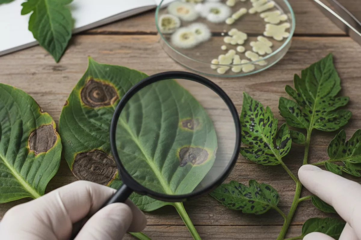
When dark leaf spots all look “about the same,” a few simple checks can quickly narrow the cause. The goal is to separate look-alike fungal leaf spot diseases from bacterial spotting and blights, then decide whether you need lab confirmation or can act confidently based on field clues.
- Start with a hand lens check (10×–20×): Look for tiny black dots (often fungal fruiting bodies) embedded in older lesions. If you see pepper-like structures arranged in the dead tissue, fungi move higher on the list. A bacterial spot usually lacks these discrete fruiting bodies and may look more water-soaked at the margins.
- Do the “water-soaked edge” test: Hold the leaf at an angle in bright light. Bacterial lesions commonly have a greasy, translucent halo early on, while many fungal lesions look drier and more papery as they expand.
- Check for angular shape limited by veins: Spots that form sharp, blocky angles (because veins act like walls) often suggest bacteria. Fungal lesions are frequently more circular or irregular and less strictly vein-bounded.
- Look for concentric rings or target patterns: A bullseye look (rings within the spot) is a classic fungal clue on many hosts. It’s not universal, but when present it’s useful.
- Try the bacterial “streaming” test: Cut a small piece from the edge of a fresh lesion (include healthy-to-diseased tissue), place it in a clear glass with clean water, and watch for cloudy threads streaming from the cut edge within 1–5 minutes. Streaming supports a bacterial cause; no streaming doesn’t rule it out, especially if the tissue is old or dried.
- Smear test for ooze (when symptoms are fresh): Gently press the lesion edge with a clean fingertip or swab. Some bacterial infections produce a sticky film that smears; many fungal spots won’t. Avoid doing this on valuable plants if you might spread pathogens—sanitize tools and hands afterward.
- Humidity response overnight: If you can isolate a leaf, place it in a sealed container with a slightly damp paper towel (not touching the leaf) for 12–24 hours at about 21–24°C (70–75°F). Fungal problems may show faint surface growth or sporulation; bacterial lesions may expand with more water-soaking. Discard the setup afterward to avoid spreading disease.
- Pattern on the plant matters: Fungal leaf spots often start on lower/older leaves and move upward with splashing water. Bacterial issues can explode after storms, overhead irrigation, or handling wet plants, sometimes showing more rapid spread across the canopy.
- Timing after weather events: A sudden jump in new lesions 2–5 days after warm, wet conditions (for example, 27–32°C / 81–90°F days with humid nights) can fit either group, but bacteria often show a sharper “overnight” change, especially with wind-driven rain.
- Rule out non-infectious lookalikes: Fertilizer burn, pesticide phytotoxicity, sunscald, and salt injury can create dark patches. These often appear more uniformly across exposed areas, may follow spray patterns, and usually lack the progressive, spreading margins seen with living pathogens.
| Clue you can observe | More consistent with fungal disease | More consistent with bacterial disease |
|---|---|---|
| Lesion texture and edge | Drier, papery center; edges less “wet” looking | Water-soaked or greasy-looking edge, especially when fresh |
| Spot shape relative to veins | Often round/irregular; not strictly vein-limited | Frequently angular; bounded by veins |
| Surface signs under 10×–20× lens | Possible tiny black dots/fruiting bodies in older lesions | Usually no fruiting bodies; may show subtle sheen/ooze when fresh |
| “Streaming” in clean water (1–5 min) | Typically negative | Often positive (cloudy threads from cut tissue) |
| Response to high humidity (12–24 hours) | May develop faint surface growth/sporulation | May enlarge with more water-soaking; sporulation absent |
| Speed of visible spread after rain/handling | Gradual increase; linked to splash dispersal | Can surge quickly after storms, overhead watering, or working plants wet |
If you still can’t tell, prioritize a fresh sample: choose leaves with early lesions (not fully dried), place them in a paper bag (not plastic), and keep them cool around 4–10°C (39–50°F) until you can get a diagnosis. Old, crispy spots are harder to interpret because both fungi and bacteria can stop showing their telltale signs once tissue is dead.
Treatment differences by disease type
Management changes a lot depending on whether you’re dealing with a fungus or bacteria. Fungal leaf spots often respond to protective sprays and better airflow, while bacterial problems are more about sanitation, keeping foliage dry, and preventing spread—because “curing” infected tissue is rarely realistic.
| Disease type | What usually helps most | What to avoid | Notes for dark leaf lesions |
|---|---|---|---|
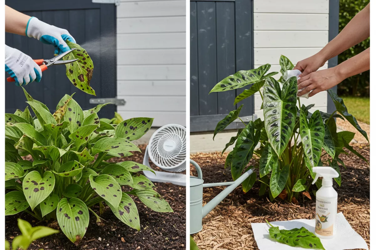
| Fungal leaf spot / blight | Remove heavily affected leaves; thin dense growth for airflow; water at the soil line; apply a labeled fungicide preventively during wet periods | Overhead watering; leaving infected debris on the soil; repeated use of the same fungicide group without rotation | Fungal lesions often expand with a visible margin or halo; stopping new infections is the goal, since existing spots won’t “heal” |
| Bacterial leaf spot / blight | Strict sanitation (discard infected leaves, don’t compost); avoid splashing water; disinfect tools; copper-based bactericides can reduce spread when used early | Handling wet plants; overhead irrigation; saving seed or cuttings from symptomatic plants | Bacterial spots can look water-soaked or greasy at first, then turn dark; focus on slowing spread rather than expecting a quick turnaround |
| Fungal rust / mildew-type issues | Increase sun and airflow; remove the worst leaves; treat early with a suitable fungicide; keep humidity down where possible | Crowding plants; high nitrogen feeding that pushes soft, dense growth; letting humidity stay high overnight | These can coexist with other leaf diseases; if you see multiple symptom styles, treat culture conditions first, then target the dominant issue |
| Mixed or uncertain cause | Start with low-risk steps: isolate plant, prune and bag damaged foliage, clean tools, switch to drip/soaker watering, and improve spacing; monitor new growth for 7–14 days (1–2 weeks) | Spraying multiple products at once; escalating to strong chemicals without confirming label suitability for the plant | If new leaves keep developing fresh dark lesions despite drier foliage and sanitation, reassess: bacteria is more likely when spread follows handling/splashing patterns |
- Timing matters: fungicides work best before or at the very first sign of spotting; bactericides are mainly suppressive and most useful early, before lesions are widespread.
- Water management is a shared priority: keep leaves dry by watering in the morning and at the base of the plant; if you must use overhead watering, do it early so foliage dries quickly.
- Pruning strategy differs slightly: for fungal problems, removing the worst leaves plus debris cleanup can noticeably slow reinfection; for bacterial issues, be more aggressive about bagging and disposing of infected material and disinfecting tools between cuts.
- Expectations: neither type typically “repairs” damaged leaf tissue—success looks like clean new growth and fewer new spots, not the disappearance of old lesions.
Common misdiagnosis mistakes
Most wrong calls happen when you rely on one clue (usually color) and ignore pattern, leaf age, and recent weather. Dark spotting can come from fungi, bacteria, sunscald, chemical splash, or even nutrient issues, so a quick glance often leads to the wrong treatment.
- Judging by “dark = fungal”: Many bacterial problems also create brown-to-black lesions. Instead of color, look for how the spot expands and whether it’s limited by leaf veins (a frequent bacterial hint) or shows concentric rings (more common with some fungi).
- Ignoring the leaf’s position and age: Spots that start on older, lower leaves after long wet periods often point to splash-dispersed pathogens. Lesions appearing first on tender new growth can suggest a different cause (including spray injury or fast-moving bacterial infections).
- Missing the “water-soaked” phase: Bacterial leaf spots often begin as greasy or translucent areas before turning dark. If you only inspect after everything has dried, you may misread the disease as a dry, papery fungal blight.
- Overweighting yellow halos: A yellow margin around a lesion can occur with bacteria, fungi, herbicide drift, and some nutrient imbalances. Treat halos as supporting evidence, not the deciding factor.
- Confusing leaf scorch with infection: Heat, wind, salt, or fertilizer burn can create dark, crisp edges that look like “blight.” Scorch usually follows a uniform pattern (tips/edges, sun-exposed side) rather than scattered lesions with distinct borders.
- Assuming fuzzy growth must be present: Many fungal leaf diseases don’t show obvious mold on the leaf surface until conditions are very humid. If you wait to see fuzz, you may delay action or mislabel a bacterial issue as “not fungal.”
- Not checking the underside of the leaf: Some pathogens sporulate or ooze more on the underside. Flip leaves and use a hand lens if you have one; surface texture and tiny fruiting bodies can change the diagnosis.
- Mixing up “shot holes”: Holes can form when dead tissue drops out, but insects can do the same. If you see clean chewing marks or frass, don’t assume a leaf-spot pathogen is responsible.
- Misreading spray damage as disease: Copper, sulfur, oils, and some soaps can cause dark speckling or larger blotches, especially if applied in strong sun or at high heat (for example, above 30°C (86°F)). A pattern that mirrors spray coverage (one side of the plant, or only the top canopy) is a big clue.
- Treating before confirming spread: A single plant with lesions after a storm or overhead watering might be mechanical injury plus opportunistic microbes. If the pattern isn’t expanding to nearby plants over 7–14 days (1–2 weeks), an aggressive disease program may be unnecessary.
- Skipping a simple “wipe test” for bacterial ooze: On very fresh lesions, gentle pressure can sometimes produce a sticky smear (more suggestive of bacteria). It’s not definitive, but it’s a useful cross-check alongside lesion shape and vein-limited patterns.
- Assuming one cause explains every symptom: It’s common to have a primary issue (like a fungal leaf spot) plus secondary stress (drought, nutrient imbalance) that changes how lesions look. When symptoms don’t fit neatly, consider a mixed scenario before choosing a control.
If you’re torn between fungal and bacterial causes, slow down and document: note recent rain/overhead irrigation, temperature swings, and whether spots are spreading plant-to-plant. A few consistent observations usually beat a “best guess” based on lesion color alone.